You must be signed in to read the rest of this article.
Registration on CDEWorld is free. Sign up today!
Forgot your password? Click Here!
Many occlusal diagnostic strategies can be burdensome, demanding extensive occlusal and temporomandibular joint (TMJ) evaluations for all patients. While this exercise may be considered ideal and necessary for an occlusal/temporomandibular disorder (TMD) rehabilitation treatment center, it may be deemed an excessive use of resources for most dental practices. The conservative use of healthcare resources is an issue in medicine, and similar paradigms must be considered for dentistry. For the busy clinician to be able to routinely perform an occlusal evaluation on every patient, the process must be methodical, incremental, and uncomplicated. The occlusal disease management system described in this article offers an approach that attempts to satisfy these requirements.
Rationale For Occlusal Disease Diagnosis
The masticatory system has three primary enemies: tooth caries, periodontal disease, and the damage caused by occlusal disease, in all of its forms. Historically, the dental profession has been focused on preventing and repairing the damage of dental caries. More recently, greater emphasis has been placed on managing periodontal diseases because knowledge has expanded for diagnosing and treating oral flora. The presence of active occlusal prematurities has been associated with the progression of periodontal diseases.1 Further, while the consequences of occlusal disease have become increasingly apparent, often the condition goes undiagnosed and untreated. Even though cervical dentin hypersensitivity and abfractive lesions are common problems seen in the dental office, many practitioners do not associate them with the underlying occlusal disease.2-4 Dentists need to be aware that the cervical stress from excessive chronic horizontal occlusal force may continue as an etiological factor for the maturation of cervical defects or restorative loss until these loading forces are removed.
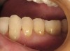
As patient demand for esthetic restorations has increased, the use of restorative materials such as porcelain and resin-based composites also has increased.5 Occlusal disease in the form of hyperocclusion, also called chronic occlusal microtrauma, is often the main reason for premature operative, esthetic, or prosthetic failure,6-9 including Class 5 restorations (Figure 1).
Occlusal Disease Education
Dentists appear reluctant to treat occlusal disease.10 This may be because occlusal education in dental schools is often minimal, and after graduation, available resources for additional training are limited. Courses offering continuing education about occlusion can be costly and time-demanding for many clinicians. Additionally, occlusion courses encourage the concepts of comprehensive and detailed occlusal/ TMD analysis for all patients. The time–intensive nature of these protocols often leads to lack of use. This may be an example of overzealous data gathering, as explained by Stohler: "Unnecessary data gathering cannot be regarded as a measure of thoroughness."11
The medical profession is keenly aware of the limited resources available to diagnose and treat patients. Although medical professionals often disagree with constraints, they generally support conservative examination methods that avoid excessive expense. Only diagnostic methods which show clear and beneficial cost-benefit ratios are suggested for use in healthcare treatment.12,13
Adding to the complexity, most occlusion courses mix occlusion and TMD. This may imply that professionals who desire to gain knowledge in occlusion must be additionally proficient in TMD diagnosis and treatment. All dentists must be occlusal experts, in that the consequences of intra-arch tooth contacts impact masticatory system function as well as affect the durability of restorative treatment. However, even though all clinicians need to have the ability to recognize and assess TMJ health or TMD, it is beyond reason that all clinicians need the expertise to treat complex TMDs. A methodical and uncomplicated diagnostic protocol for occlusal disease would seemingly help the profession confront masticatory system imbalance.
Occlusal Disease Management System
The Occlusal Disease Management System was designed to simplify the diagnostic process, and uses an incremental system of stages to classify the severity of occlusal/TMD pathology.2,14 When extensive restorative and esthetic dentistry is anticipated, this system may be used with the Dento-Facial Esthetic Diagnosis (DFED) system.15,16 The DFED system records the patient's goals, personality, preferences, and conditions that may impact patient acceptance of treatment. It then blends clinical determinants with these personal elements, as well as the 25 parameters of dentofacial esthetic design.
Stage 1: Occlusal and TMJ Screening
Every comprehensive dental examination must include a basic occlusal and TMJ screening, but for this to take place routinely, it should not be time-consuming. Discovery questions and clinical evaluation are intended to screen for signs and/or symptoms of occlusal disease. This data may aid in diagnosing either current or prior conditions that impact the prognosis for minor or extensive restorative treatment. If progressive occlusal disease or temporomandibular pathology is detected, then the patient should be offered a more complete occlusal and TMJ examination. This service may involve a separate fee and a subsequent appointment, because it requires an extensive occlusal or TMJ examination and mounted casts.
Some patients are reluctant to address occlusal or TMD issues. These patients must be advised of the consequences, and their reluctance should be recorded. By performing an initial occlusal/TMJ screening, the clinician gathers information that could impact patients' insight into masticatory system requirements for healthy function, which may lead, in time, to a change in these patients' point of view of the recommended treatment. A clinician should use caution when considering even minimal restorative dentistry when a patient avoids a comprehensive examination of the masticatory system, or refuses to acknowledge his or her occlusal condition. In some extreme cases, a refusal to go forward with treatment for this type of patient may be indicated to avoid a failure in expectations.
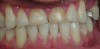
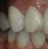
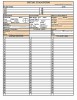
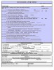
The signs and symptoms of occlusal disease are pathological occlusal tooth wear (Figure 2), fractures of teeth/ restorations (Figure 3), hypersensitivity of teeth during mastication, cervical dentin hypersensitivity,3,4 tooth hypermobility,1 fremitus,1,2 abfractions,2,3,5,6,17 vertical bone loss or localized bone destruction (secondary to periodontal disease),1 and masticatory muscle or TMJ pain.6,18,19 Each dentist's screening system must allow him or her to determine if the patient presents any of these signs or symptoms. As shown in Figure 4, the initial examination form should include a section dedicated to occlusal and TMJ screening. Simple "yes" or "no" answers may be sufficient for some questions. Stage 1 evaluation is designed to detect signs or symptoms of occlusal disease that predicate the need for a Stage 2 occlusal and TMJ examination. Because the diagnosis and treatment of occlusal disease requires mounted casts and a more extensive occlusal or TMJ examination, the clinician should refrain from making diagnostic decisions related to treatment at this time.
Enough information may be gathered in a few minutes to determine if occlusal disease or instability exists within the masticatory system that warrants further investigation. If any of the discovery questions receive a positive answer, and especially if the patient answers that he or she experiences the symptom with an increased level of severity, a Stage 2 occlusal and TMJ examination is recommended. When multiple positive answers are present, then the patient should be advised that he or she presents signs/symptoms of either occlusal disease or TMD. Patients often are surprised when there is discussion about occlusal disease because only a limited number of clinicians have the background or inclination to share this information with patients.10 It seems to the authors that masticatory system diagnosis is the key to effective treatment. The consequences of nontreatment, according to current literature, should be presented to the patient. Proper education and motivation are prime factors in attaining clinical success for both restorative and esthetic treatment. Freedom from active occlusal disease or TMD pathology must exist before instigating a dental therapy that relates to function of the masticatory system.
Occlusal or TMD therapy should not be initiated until after a Stage 2 examination with mounted study casts and a more thorough evaluation. The exception to this rule is the fabrication of a night guard (NG), which is the most basic preventive measure dentists can provide patients with occlusal disease. Often, patients will accept an NG even if they refuse more advanced evaluation of occlusal or temporomandibular health. An NG should be constructed free of lateral posterior interferences with cuspid-rise or flat-plane working cusp contacts to reduce neurosensory input to the muscles of mastication from tooth contacts. It is beyond the scope of this article to elaborate on the topic of appliance therapy. The NG should not be fabricated as a therapeutic splint that alters the anterior/superior condyle position in the glenoid fossa defined by centric relation (CR) (as discussed in Stage 2). A more precise TMJ examination (Stage 3), as well as appropriate radiographic analysis, is indicated before appliance therapy for joint pathology. An NG is a preventive reversible appliance. Patients need to be informed that in the rare event an NG causes pain, they need to discontinue its use, and that they will need a more comprehensive diagnosis before further treatment.
Stage 2: Occlusal and TMJ Examination
After the initial dental examination and occlusal/TMJ screening, patients who are prognostic risks because of the presence of occlusal or temporomandibular pathology are advised to have an occlusal and TMJ examination using an examination form, such as the one in Figure 5. This examination involves a comprehensive occlusal evaluation and a Stage 2 TMJ screening. The authors do not recommend an advanced TMD evaluation unless the clinician has sufficient training to treat complex joint pathology. The three primary objectives of this examination are: to understand the etiology of the occlusal condition; to determine if occlusal therapy is indicated/feasible; and to determine if pathology exists within the TMJs that may contraindicate restorative dentistry and require treatment. If an advanced or complex TMD condition is diagnosed, then referral to a TMD specialist may be indicated. The good news is that patients exhibiting more severe TMD are a small percentage of the population and may be identified during this Stage 2 examination.
Uncovering occlusal or TMJ disease is a process of discovery for the dentist and the patient alike. The upper portion of the examination form should be a patient questionnaire designed to help uncover potential signs/symptoms of occlusal or temporomandibular disease. Pathologic temporomandibular signs or symptoms may include: headaches, pain in the TMJs or muscles of mastication, joint noise on mandibular opening/closing, limited mandibular opening, grating or popping noises within joints, and difficulty with the mastication of foods. Familial arthritic conditions often are shared by subsequent generations, and because the TMJs are the most active joints in the body, arthritic pathology present in other joints also may appear in these joints. Pain in the muscles of mastication, and often within neck or shoulder muscles, may be a result of posturing the mandible to avoid deflective tooth contacts. These components relate to one another in health and during the events of pathology. Macrotrauma (eg, an automobile accident) traumatizes bone, muscular soft tissue, and dental components at a moment in time. Chronic occlusal microtrauma affects the masticatory system, causing occlusal disease. Both clenching and/or parafunctional activity may result in stress or pain to masticatory muscles. Therefore, the form should include questions that can help the clinician and patient determine if connections exist between signs/symptoms of TMD and emotional/stressful events.
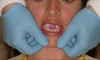
Stage 2 evaluation includes a comprehensive clinical occlusal examination, which should be documented. The clinical examination assists in the determination of pathology within the masticatory system during the interaction between TMJs, muscles of mastication, and interdigitation of teeth. Neurosensory connections exist between the masseter/ temporalis muscles and dental pulp/periodontal proprioceptors to regulate the contractility by these muscles of mastication.20-22 Premature contacts on teeth during function have been found to produce alterations in the contraction-release cycles by the muscles that position the mandible. These premature contacts of teeth or an accommodation by masticatory muscles may spell disaster to restorative or esthetic treatment.20-22 The ability to place the mandible in CR during the clinical examination is useful for evaluating joint health. CR is a joint position where the condylar head is in the most anterior/superior position within the glenoid fossa and the meniscus is centered between the condyle head and bone of the fossa. The technique of bimanual manipulation was introduced by Peter Dawson as a method for locating CR.23 Using an anterior deprogrammer for 10 to 20 minutes may be useful to facilitate bimanual manipulation in some patients (Figure 6). A load test, also described by Dawson, provides information about joint inflammation and meniscus position in CR.23 Centric occlusion (CO) is the maximum intercuspation of teeth in the temporomandibular position of CR.
Dental horizontal incline deflections have been implicated in the formation, over time, of cervical defects known as abfractions.3,4 The presence of abfractions detected during the examination must be noted, in that they may represent either prior or current occlusal prematurities. Noncarious cervical lesions may include hard-tissue lesions formed as a result of stress (abfractions), friction (toothbrush/dentifrice abrasions or occlusal/incisal attrition), or corrosion (formerly termed chemical degradation or erosion).24 The diagnosis of cervical dentin hypersensitivity appears to represent the early or active expression of the pulp when occlusal deflection produces microfracture of hydroxyapatite during the formative stage of an abfraction.4
The air-indexing method objectively detects the presence or absence of a threshold patient response to an air stimulus for the diagnosis of cervical dentin hypersensitivity. To investigate a response to this stimulus, a minor puff of room-temperature air is directed toward the cementoenamel junction at a 45° angle to the long axis of a test tooth for 0.5 to 1 second at a distance of approximately 0.5 cm.25 A positive air index verified at a subsequent visit 7 to 10 days after the initial detection of an air response suggests the presence of active abfractive stress from chronic occlusal microtrauma.3 Corrosive agents may accelerate the mechanism of cervical stress by opening root dentin tubules, which reduces the threshold for the mechanoreceptor response of cervical dentin hypersensitivity.
Because it is unlikely that all exposure of human teeth to corrosive agents can be removed, the presence of cervical dentin hypersensitivity should not always be interpreted as an objective sign/symptom of occlusal disease.25 In general, when less than five or six teeth have been diagnosed with verified cervical dentin hypersensitivity, the primary etiology may be from chronic occlusal microtrauma. An individual diagnosed with greater than six teeth with the presence of cervical dentin hypersensitivity may have a primary etiologic factor of corrosion. The interplay between stress, corrosion, and friction seems responsible for the formation of noncarious cervical lesions.
Findings of multiple tooth fractures over time in the patient history suggest that occlusal intervention may be indicated. Tooth contacts may be evaluated with the help of occlusal wax indicators; articulating papers; or a novel technology, the T-Scan® III computerized occlusal analysis system (Tekscan, Inc, www.tekscan.com). Freedom from lateral incline contacts by posterior teeth during excursive movements has been suggested to reduce undesirable horizontal interferences.26,27 The authors suggest that a comprehensive Stage 2 occlusal examination should result in definitive treatment only when the patient's acceptance and clinician's opinion correspond in the effort toward maximal dental health.
A trained dental assistant may take high-quality impressions of the patient's dentition, as well as oral photographs. An accurately mounted set of study casts may be obtained only with an occlusal CR record (CR bite), face bow record, and properly poured stone. This study cast mounting allows for an evaluation of CR deflections and a trial equilibration to gain a deeper understanding for possible therapy. Armed with the completed forms, study cast analysis, clinical evaluation findings, as well as oral photographs, the clinician has enough information to complete the occlusal and TMJ examination. This information should be presented to the patient at a treatment presentation appointment.
The scope of this article does not include a detailed explanation of treatment; nevertheless, the resolution of occlusal deflections should occur before the completion of either restorative or esthetic treatment plans. Minor tooth deflections that reduce the maximum intercuspation in CR should be equilibrated, if possible, after the study cast is analyzed and a trial equilibration performed. The prognosis for the restoration of cervical defects is, in large part, dependent on the presence of excessive occlusal loading. Psychological stability or the lack thereof has been correlated to the risk of active bruxism. The intent of Stage 2 examination is to determine which occlusal/temporomandibular pathologies warrant treatment and which conditions are not anticipated to alter the esthetic or restorative prognosis of treatment.
Stage 3: Advanced Occlusal and TMJ Examination
Stage 3 examinations should provide the clinician with the information to either treat or refer patients diagnosed with TMD or joint disease. Clinicians who are providing treatment that involves incisal edges or occlusal surfaces of teeth should have the greatest knowledge about occlusion and how to treat occlusal disease, but they do not need to perform advance diagnosis or to treat TMD. The arena of TMD diagnosis and treatment has become more specialized with a multiplicity of professionals contributing to diagnosis as well as to treatment. Radiographic analysis may include computed tomography and/or magnetic resonance imaging. More recent imaging techniques include 3-dimensional imaging of the TMJ. If the patient is hesitant to participate in the comprehensive examination necessary to evaluate occlusal, muscular, or temporomandibular health, then these details should be noted in the patient record. If the clinician feels overwhelmed by the presence of TMD or joint disease, it is appropriate to refer the patient to a specialist to ease the burden of treatment responsibility. Restorative or esthetic treatment must be delayed until a margin of safety for the prognosis of treatment is achieved.
Conclusion
Restorative clinicians can and should be experts in occlusion with the ability to treat occlusal disease. Helping patients toward masticatory system health or the recognition of pathology is a professional service that may enhance the prognosis for restorative and/or esthetic treatment. This article showcased an approach—The Occlusal Disease Management System—that is methodical, incremental, and uncomplicated: all indispensable factors for the busy clinician who wishes to routinely perform an occlusal evaluation of all patients.
References
1. Harrel SK, Nunn MP, Hallmon WW. Is there an association between occlusion and periodontal destruction? Yes—occlusal forces can contribute to periodontal destruction. J Am Dent Assoc. 2006;137(10):1380-1384.
2. Ruiz JL. Achieving longevity in esthetic dentistry by the proper diagnosis and management of occlusal disease. Contemporary Esthetics. 2007;11(6):24-29.
3. Coleman TA, Grippo JO, Kinderknecht KE. Cervical dentin hypersensitivity. Part III. Resolution following occlusal equilibration. Quintessence Int. 2003:34(6):427-434.
4. Coleman TA, Grippo JO, Kinderknecht KE. Cervical dentin hypersensitivity. Part II. Associations with abfractive lesions. Quintessence Int. 2000;31(7):466-473.
5. Ruiz JL. The psychology of a smile. Journal of Cosmetic Dentistry. 2003;19(1):58-59.
6. Ruiz JL. Achieving optimal esthetics in a patient with severe trauma: using a multidisciplinary approach and an all-ceramic fixed partial denture. J Esthet Restor Dent. 2005;17(5):285-291.
7. Christensen GJ. A look at state-of-the-art tooth-colored inlays and onlays. J Am Dent Assoc. 1992;123(9):66-70.
8. Ruiz JL, Christensen GJ. Rationale for the utilization of bonded nonmetal onlays as an alternative to PFM crowns. Dent Today. 2006;25(9):80-83.
9. Tyas MJ. The class V lesion—aetiology and restoration. Aust Dent J. 1995;40(3):167-170.
10. Christensen GJ. Abnormal occlusion conditions: a forgotten part of dentistry. J Am Dent Assoc. 1995;126(12):1667-1668. 11.McNeill C, ed. Science and Practice of Occlusion. Chicago, IL: Quintessence Books; 1997:294-296.
12. Udvarhelyi IS, Colditz GA, Rai A, et al. Cost-effectiveness and cost-benefit analysis in the medical literature. Are the methods being used correctly? Ann Intern Med. 1992;116(3):238-244.
13. Doubilet DP, Weinstein MC, McNeil BJ. Use and misuse of the term cost effective in medicine. N Engl J Med. 1986;314(4): 253-256.
14.Ruiz JL. Occlusal disease: restorative consequences and patient education. Dent Today. 2007 26(9):90-95.
15. Ruiz JL. A systematic approach to dento-facial smile evaluation using digital photography and a new photographic view. Dent Today. 2006;25(4):82-85.
16. Ruiz JL. Achieving predictable, beautiful smiles using a dento-facial diagnostic system. Compend Contin Educ Dent. 2007;28(1):50-55.
17. Grippo JO. Abfractions: a new classification of hard tissue lesions of teeth. J Esthet Dent. 1991;3(1):14-19.
18. Gremillion HA. The relationship between occlusion and TMD: an evidence-based discussion. J Evid Based Dent Pract. 2006;6(1):43-47.
19. Sipila K, Ensio K, Henhela H, et al. Occlusal characteristics in subjects with facial pain compared to pain-free control group. Cranio. 2006;24(4):245-251.
20. Sharav Y, Mc Grath PA, Dubner R. Masseter inhibitory periods and sensations evoked by electrical tooth pulp stimulation in patients with oral-facial pain and mandibular dysfunction. Arch Oral Biol. 1982;27(4):305-310.
21. Holmgren R, Sheikholeslam H, Riise C. The effects of an occlusal splint on the electromyographic activities of the temporal and masseter muscles during maximal clenching in patients with a habit of nocturnal bruxism and signs and symptoms of craniomandibular disorders. J Oral Rehab. 1990;17(5):447-459.
22. Suganuma T, Ono Y, Shinya A, et al. The effect of bruxism on periodontal sensation in the molar region: a pilot study. J Prosthet Dent. 2007;98(1):30-35.
23. Dawson PE. Evaluation, Diagnosis, and Treatment of Occlusal Problems. 2nd ed. St. Louis, MO: CV Mosby; 1989:28-55,434-441.
24. Grippo JO, Simring M, Schreiner S. Attrition, abrasion, corrosion, and abfraction revisited [published erratum in J Am Dent Assoc. 2004;135(10):1376.]. J Am Dent Assoc. 2004;135(8):1109-1118.
25. Coleman TA, Kinderknecht KE. Cervical dentin hypersensitivity. Part I: The air indexing method. Quintessence Int. 2000; 31(7):461-465.
26. Williamson EH, Lundquist DO. Anterior guidance: its effect on electromyographic activity of the temporal and masseter muscles. J Prosthet Dent. 1983;49(6):816-823.
27. Kerstein RB, Wright NR. Electromyographic and computer analyses of patients suffering from chronic myofascial pain-dysfunction syndrome: before and after treatment with immediate complete anterior guidance development. J Prosthet Dent. 1991;66(5):677-686.
About the Authors
Jose-Luis Ruiz, DDS
Course Director
University of Southern California Advanced Esthetic Dentistry Continuum
Los Angeles, California
Clinical Instructor
University of Southern California
Los Angeles, California
Private Practice
Burbank, California
Thomas A. Coleman, DDS
Private Practice
Brandon, Vermont